
Tardigrade observation guide
How to find a tardigrade under a microscope
Finding a water bear is part careful sampling and part patient scanning; the right low-power workflow matters more than extreme magnification.
Start with a realistic search image
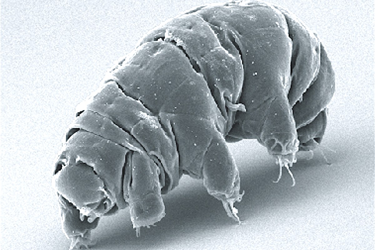
A tardigrade under a microscope has a compact, usually translucent body and four pairs of short legs. The rear pair points backward, and small claws may be visible when focus and contrast are favorable. Its deliberate crawl inspired the name “water bear,” but movement varies with the animal, temperature, water depth, and condition of the sample.
University of Florida IFAS Extension describes tardigrades as small invertebrates ranging roughly from 0.1 to 1 millimeter long. That makes many individuals visible with ordinary light microscopy, but they can be difficult to locate among moss fragments, nematodes, rotifers, and other debris.
Tardigrades are not guaranteed in every sample. Collect several small samples from suitable places and treat an unsuccessful search as information rather than a reason to jump immediately to maximum power.
Collect moss or lichen responsibly
Many terrestrial tardigrades live in the thin film of moisture around mosses, lichens, leaf litter, and similar habitats. Cornell's tardigrade classroom lab uses a simple collection and extraction activity based on moss and lichen.
Before collecting:
- get permission from the property owner or site manager;
- avoid protected land or protected species unless collection is explicitly allowed;
- take a small pinch rather than stripping a patch;
- choose material within safe reach instead of climbing;
- keep samples from different locations in separate labeled paper bags.
Dry moss from a tree trunk, rock, wall, or roof edge can still be worth examining because some tardigrades enter a contracted, dehydrated state when liquid water is unavailable. This survival response does not make them indestructible. Heat, abrupt chemical exposure, physical pressure, and prolonged drying can still damage them.
Record the location, surface, date, and whether the moss was dry or damp. Those notes make it possible to compare which microhabitats produced results.
Extract the animals into water
Use a shallow, clean dish, a dropper or pipette, and rainwater, distilled water, or another water source specified by your instructor's protocol. Place a small amount of moss or lichen in the dish and add enough water to wet it thoroughly.
Educational collection protocols vary in soaking time. A practical first attempt is several hours; some established procedures allow the material to soak for up to about a day. Keep the dish covered loosely against dust and away from heat or direct sun.
After soaking:
- Remove or lift the plant material with clean tweezers.
- Gently squeeze or agitate it over the dish so trapped water and fine debris fall out.
- Let coarse debris settle briefly.
- Draw liquid from near the settled material with a dropper.
A stereo microscope is convenient for searching the shallow dish at low magnification and transferring a located animal. If only a compound microscope is available, make multiple small wet mounts and scan them systematically. Do not place a deep, debris-filled dish on a stage that is designed for glass slides.
Prepare a searchable wet mount
Place one small drop of extracted water in the center of a clean slide. If the drop is crowded with moss pieces, choose a clearer portion or dilute it with a small amount of clean water. Lower a cover slip at an angle to reduce air bubbles.
Do not press on the cover slip. Pressure can injure an animal and also move liquid unpredictably across the slide. If large debris prevents the cover slip from resting normally, remake the slide with less material.
Start with the lowest-power objective. With a 10X eyepiece and 4X objective, 40X total magnification provides a wide field for searching. After centering a possible tardigrade, a 10X objective provides 100X total magnification with the same eyepiece and may reveal the body and legs more clearly. A 40X objective can show smaller features in favorable preparations, but the narrow field and shallow working distance make a moving animal harder to follow.
The magnification guide explains how eyepiece and objective powers combine and why a larger number does not guarantee more resolved detail. Oil immersion is unnecessary for this activity; never place oil on an objective that is not designed for it.
Scan instead of searching at random
Focus first on a sharp piece of debris near one edge of the cover slip. Use coarse focus only at low power, then fine focus. Adjust the transmitted light so transparent objects have useful contrast.
Move the slide through overlapping rows:
- Scan from left to right across the upper part of the cover slip.
- Move down by slightly less than one field width.
- Scan back from right to left.
- Repeat until the entire wet area has been covered.
Pay special attention to fine debris and the edges of moss fragments. Tardigrades may crawl slowly enough to resemble a stationary clump at first, so pause and watch likely shapes for several seconds.
If the subject disappears after changing objectives, return to low power, re-center, and focus again. Never force a focus control or allow an objective to strike the slide.
Distinguish a tardigrade from common look-alikes
Look for a combination of traits rather than one vague resemblance. Arizona State University's anatomy guide highlights the eight legs as a practical distinguishing feature.
| Observation | Tardigrade | Possible look-alike |
|---|---|---|
| Body | Plump, segmented-looking outline | Fiber or plant fragment may be rigid and irregular |
| Appendages | Four pairs of short legs | Nematodes and fibers have no legs |
| Movement | Slow crawl or body contraction | Nematodes often thrash; debris drifts with currents |
| Rear end | Fourth leg pair points backward | Rotifers lack four pairs of stubby legs |
| Feet | Claws may be visible with sufficient detail | Air bubbles have bright smooth edges but no anatomy |
Do not assign a species from a casual brightfield view. Species-level identification can depend on fine structures, eggs, claws, mouthparts, and specialist taxonomic methods. “Tardigrade observed” is an appropriate record when the defining body plan is clear.
Capture useful photos or video
Short video is often more informative than a single frame because the crawl, leg motion, and body contractions help distinguish the animal from debris. Focus the microscope before attaching a phone, align the active camera over the eyepiece, and keep digital zoom modest.
The Wevision M2 includes a smartphone adapter as well as a dropper and tweezers in its kit, which can support this observation workflow. The M1 and M2 both provide low-to-higher objective progressions; compare their documented controls and included items on the microscope comparison page. The smartphone microscope photography guide explains alignment, exposure, vibration control, and recordkeeping.
Note the sample source, soak time, objective, and whether the image was cropped. Do not infer an animal's exact size from an uncalibrated photo.
Protect the specimen and finish cleanly
Limit unnecessary time under bright illumination, which can warm a small wet mount and accelerate evaporation. If the cover-slip edge begins drying, end the observation rather than pressing on the glass or flooding the stage.
Follow classroom or local instructions for sample disposal. Do not release material into a habitat different from the collection site. Clean reusable slides appropriately, place broken cover slips in a designated sharps container, wipe the work area, and wash your hands.
Finding a tardigrade rewards a methodical process: collect lightly, soak patiently, prepare a thin mount, scan at low power, and confirm the eight-legged body plan before increasing magnification. Even a slide without a water bear can reveal rotifers, nematodes, algae, and other members of the same miniature habitat.
Sources
Explore Wevision microscopes
Find the right microscope for your next observation
Visit the Wevision web store to explore current microscope models for learning, laboratories, classrooms, and hands-on discovery.
Visit the Wevision web store