
Plant cell observation guide
Elodea under a microscope: find chloroplasts and watch them move
A thin Elodea leaf lets you see green chloroplasts in living plant cells with a compound microscope. A drop of water and a coverslip are enough to prepare the leaf.
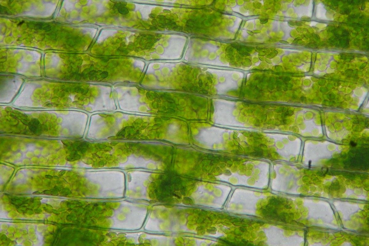
Choose a cell you can follow
Make a drawing of one Elodea cell. Mark the cell wall and several green chloroplasts. Then watch the same cell for one minute and record whether a chloroplast changes position along the wall. The drawing and timed note are your results. You can make both even when you see no clear movement.
Elodea is a freshwater plant. Its chloroplasts contain chlorophyll, the green pigment that absorbs light for photosynthesis. When the fluid and other material inside a living cell flow, they can carry the chloroplasts along. This flow is called cytoplasmic streaming. The green color can help you follow a chloroplast while the cell wall gives you a fixed point for comparison.
Gather a leaf and prepare the slide
Use a fresh Elodea sprig from a classroom or aquarium supply that is permitted in your area. Keep it in its container until you need a leaf. If you have only a prepared slide, use it to draw the walls and chloroplasts. Use the Exploratorium lesson in the sources to see recorded examples of movement.
For a fresh mount, collect these items.
- A compound microscope with a light below the stage.
- A clean slide, a coverslip, a dropper, and blunt tweezers.
- A small Elodea leaf and water from its container.
- A notebook, a pencil, and a timer.
- Place one small drop of water in the middle of the slide.
- Use the tweezers to lift one small leaf from the sprig. Hold it near its base.
- Lay the leaf flat in the drop. Check that it fits under the coverslip without folding.
- Rest an edge of the coverslip next to the drop. Lower it slowly over the leaf.
- Blot excess water at the edge of the coverslip. Dry the underside of the slide before you put it on the stage.
The guide to microscope slide preparation shows how to lower a coverslip and correct common problems. Use a clean slide if the first one has a large bubble over the leaf or if the coverslip rocks.
Find the cell walls and chloroplasts
Start with your lowest-power objective, the lens above the slide. Find the leaf outline and move toward a flat area near the tip or outer edge. The thicker middle of the leaf can put several layers in the same view. Look for a patch where you can follow the walls around one whole cell.
Center the patch and switch to a 10X objective if you have one. With a 10X eyepiece, this gives 100X total magnification. Look for a grid of walls with green dots or ovals inside it. If you can follow a chloroplast at this power, use this view for your timed observation.
For a closer view, use a dry 40X objective if your microscope and slide allow it. Check the gap from the side as you change objectives. Follow the microscope instructions and use fine focus at high power. Keep the lens clear of the coverslip. Adjust fine focus slightly to bring the green spots and nearby wall into view.
Draw the outlines you can follow. Mark the green chloroplasts that you see. If the center looks clear, label it as a clear area in your drawing. A large compartment called the central vacuole takes up much of the cell, but its boundary can be difficult to see in this mount.
Check for movement inside one cell
- Choose one green chloroplast near a cell corner or where two walls meet.
- Leave the stage and focus controls still. Start the timer at zero.
- Watch for up to 60 seconds. Compare the chloroplast position with the same wall or corner.
- Record its path if you can follow it. If you lose track of it, write down when that occurred.
- Repeat with a second cell. Use the same observation time and power.
If the wall and green spots move together across the field, wait for the slide to settle. Use the wall as a fixed point. When a chloroplast follows the inside of the wall while the wall stays fixed, the movement is consistent with cytoplasmic streaming.
Use a brief video if it is hard to follow the movement by eye. The phone photography guide shows how to hold the camera steady. Keep a wall or corner visible in the frame so that you can compare positions during playback.
| Record | Example |
|---|---|
| Sample and location | Fresh Elodea leaf, thin area near the tip |
| Magnification | 10X eyepiece and 40X objective, 400X total |
| Fixed point | Corner where two cell walls meet |
| Movement | One green dot moved along the long wall during 60 seconds |
| Repeat | No clear movement in the second cell during 60 seconds |
Finish the record and clean up
If you can see walls but the green spots look blurred, return to lower power. Choose a thinner patch and focus again. If the leaf dries, folds, or drifts, make a new mount with a fresh leaf. A second mount also gives you another place to look when the first two cells show no clear movement.
Compare your drawing with the onion cell activity. Look for clear walls in both samples and record which sample showed green chloroplasts. Keep the specimen name, magnification, and observation time with your notes.
Remove the slide and clean the tools. Follow local directions for disposal of aquarium plants and water. Keep the leaf, fragments, and water out of natural waterways. Wash your hands after the activity.
Sources
Explore Wevision microscopes
Find the right microscope for your next observation
Visit the Wevision web store to explore current microscope models for learning, laboratories, classrooms, and hands-on discovery.
Visit the Wevision web store